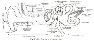
The human ear, like those of other mammals, contains sense organs that serve two distinct functions: hearing and postural balance and coordination of head and eye movements. The ear is divided into three distinct parts: the outer, middle, and inner ear. The visible portion of the ear, known as the auricle or pinna, projects from the side of the head, and the short external auditory canal, the inner end of which is closed by the tympanic membrane, also known as the eardrum. The outer ear’s function is to collect sound waves and direct them to the tympanic membrane. The temporal bone contains a narrow air-filled cavity called the middle ear.
Ear – diagram
The ear is an organ that allows humans to hear and, in mammals, to maintain body balance through the use of the vestibular system. The ear in mammals is typically divided into three parts: the outer ear, the middle ear, and the inner ear. The outer ear is the most visible of these three parts. The pinna and the ear canal are both components of the outer ear. Due to the fact that the outer ear is the only visible portion of the ear in most animals, the term “ear” is frequently used to refer to only the external portion of the ear. The tympanic cavity, as well as the three ossicles, are all located in the middle ear.

Outer part of ear
The outer ear can be found in a variety of shapes and sizes. This structure contributes to the individuality of each of us. The auricle or pinna is the medical term for the outer ear.The cartilage and skin that make up the outer ear. The outer ear is divided into three sections: the tragus, helix, and lobule.
The Ear Canal
The ear canal extends from the outer ear to the eardrum. The canal measures about an inch in length. The ear canal’s skin is extremely sensitive to pain and pressure. The outer one-third of the canal beneath the skin is cartilage, while the inner two-thirds is bone.
The Eardrum
The eardrum is approximately the size of a dime and is the same size in a newborn baby as it is in an adult. The tympanic membrane is the medical term for the eardrum. The eardrum is a gray membrane that is transparent. The middle ear bone is attached to the center of the drum (the malleus).
The Middle Ear
The middle ear is the space inside the eardrum. The malleus, incus, and stapes are three of the smallest bones in the body, located in the middle ear. These bones are also referred to as the hammer, anvil, and stirrup. The middle ear ossicles are the medical term for all three bones together.
The Ossicles
The malleus, incus, and stapes are three tiny, remarkable bones that are located near the base of the skull. As a group, they are sometimes referred to as “ossicles,” which comes from the root word “osseo,” which means “bone.”
When the tympanic membrane moves, they vibrate in response, and their shape is precisely calculated so that the vibrations are transmitted and focused, allowing them to be heard even more clearly.
These bones make contact with the eardrum, also known as the tympanic membrane, which is located on the outside of the middle ear. This causes the vibrations to be transmitted through their specially-shaped bone structures, which ultimately leads to the oval window.
How to get rid of vestibular vertigo
Vestibular Vertigo is a symptom, not a disease. It is the sensation that you or your surroundings are moving or spinning.This sensation may be barely perceptible, or it may be so intense that you struggle to maintain your balance and perform daily tasks.
Vertigo attacks can come on suddenly and last only a few seconds, or they can last much longer. If you have extreme vertigo, your illnesses may be constant and last for so many days, making normal life extremely difficult.
Treatment
The treatment of dizziness and vertigo may include medication, physical therapy, and psychotherapy; in some cases, surgical intervention may be required in rare circumstances. In order to begin treatment, it is important to inform the patient that the prognosis is generally favourable: many of these conditions have a favourable spontaneous course, both because peripheral vestibular dysfunction tends to improve and because asymmetrical peripheral vestibular tone is compensated for by central vestibular compensation.
Conclusion
This ossicular chain transports sound from the eardrum to the inner ear, which has been referred to as the labyrinth since the time of Galen (2nd century CE). It is a complex network of fluid-filled passages and dental problems located deep within the temporal bone’s rock-hard petrous portion. The inner ear is divided into two functional units: the vestibular apparatus, which includes the vestibule and semicircular canals and houses the sensory organs of postural balance, and the snail-shell-like cochlea, which houses the sensory organ of hearing. These sensory organs are highly specialised terminations of the eighth cranial nerve, also known as the vestibulocochlear nerve.
 Profile
 Settings
 Refer your friends
 Sign out


