 Profile
 Settings
 Refer your friends
 Sign out


The human ear is a hearing and equilibrium organ that detects and analyses sound through transduction (or the conversion of sound waves to electrochemical impulses) and maintains a feeling of balance (equilibrium).
The ear is separated into three regions that communicate with one another: the external, middle, and inner ears. While the external and middle ears are primarily involved with sound transmission, the inner ear houses the cochlea – frequently referred to as the organ of hearing – as well as the body’s balance organ.
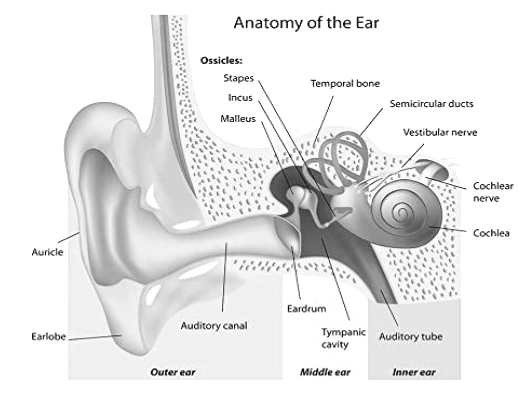
External Ear
The external ear canal, also known as the external auditory canal or external auditory meatus (EAM), is approximately 2.5cm long and coated with skin. The outer part of the pinna is cartilage; the skin that lines this cartilage contains hair follicles and wax-producing glands that are a combination of ceruminous and sebaceous glands.
The sebaceous glands discharge an oily substance called sebum into the hair follicles’ root canals, whereas the ceruminous glands are modified apocrine sweat glands that enter into the hair follicles’ base and generate a moist white secretion that darkens and thickens as it dries, becoming sticky. Ceruminous glands and hair follicles are predicted to form when the foetus is approximately five months old.
Unlike the skin on the rest of the body, which grows from the basal layers to the surface, the skin in the external ear canal migrates from the tympanic membrane to the entrance; the rate of migration differs across individuals. The shedding of migratory skin combines with gland secretions to generate wax; any accumulation in the external ear canal – such as dead skin cells, dust, debris, shed hair, or wax – migrates naturally out into the conchal bowl. This is a continuous movement from the eardrum’s centre to its periphery, then up and out of the external ear canal.
Because the glands are only present in the outer part of the external ear canal, wax should be seen only in this area. Short hairs are placed above the glands and are projected toward the canal entrance to aid in the migration of wax out of the ear canal; they also protect the ear canal by trapping dust and debris and preventing foreign objects from entering the external ear canal. Wax is somewhat acidic and protects the external ear canal from bacterial and fungal infection; hairs pull the wax slightly away from the skin in the outer third of the canal, preventing skin irritation.
The temporal bone houses the inner two-thirds of the ear canal, with the skin securely clinging to it; the diameter of the ear canal varies between individuals and races. The ear canal proceeds downward and slightly forward into the skull, narrowing and bending somewhat at the intersection of the skull’s cartilage and bone. The tympanic membrane (eardrum) is located at the end of the external ear canal. It is slanted, creating a recess in the front wall of the ear canal. Adults must raise their pinna upwards and outwards (children must move their pinna downwards and rearward) in order to view down the canal to the tympanic membrane.
The tympanic membrane separates the external and middle ears; it is slightly oval in shape and measures roughly 9-10mm in diameter at its widest point. The pars tensa comprises the majority of this membrane and is located at the tympanic membrane’s lower end. It is made up of three layers:
- Epithelium that runs parallel to the external ear canal
- Middle layer of fibrous tissue
- Mucosal membrane that lines the whole middle ear and upper respiratory tract
The pars flaccida is a triangular section located above the pars tensa that is devoid of fibrous tissue. This increases its susceptibility to cholesteatoma, an ear condition characterised by dead skin accumulating in the middle ear, resulting in discharge and hearing loss. This can cause structural deterioration in and around the middle ear, necessitating surgical removal. It is either congenital or develops as a result of recurrent ear infections.
The tympanic membrane is thickened at its outer rim, which is referred to as the annulus. The malleus, the first of three middle ear bones, is linked at its tip to the umbo, or membrane, in the centre of the ear. The malleus handle, the most distinctive feature of the tympanic membrane, is located between the fibrous and mucosal layers of the pars tensa and is joined at both the umbo and lateral processes. Due to the membrane’s slight concavity, shining a light on it during otoscopy (examination of the external ear canal and tympanic membrane with an otoscope) reveals a triangle of light toward the front and lower aspect; this is referred to as the light reflex and can assist the viewer in orienting the membrane.
Middle Ear
The middle ear is uneven in form and mucosa-lined. Although it is frequently referred to as an air-filled region, it is actually filled with nitrogen-rich gas.
The roof of the middle ear is separated from the meninges and temporal lobe of the brain by a thin bone plate called the tegmen tympani. Its floor is formed by the jugular fossa’s roof and is located adjacent to the internal jugular vein and internal carotid artery. Due to the proximity of the vein and artery, some persons complain of hearing blood pumping in their ear.
The eustachian tube is located on the anterior wall of the middle ear. This cartilage-and-bone tube connects the middle ear to the nasopharynx in the post-nasal region.
Inner Ear
The inner ear consists of:
- The cochlea (organ of hearing)
- The peripheral vestibular apparatus (organ of body balance)
Cochlea
The cochlea is a thick, snail-like structure that lies horizontally and contains the organ of Corti. Its spiral channel is 29mm to 40mm in length and is separated into three compartments by bone and membrane walls. The upper (scala tympani) and bottom (scala vestibuli) compartments are filled with perilymph, while the middle (scala media) compartment is filled with endolymph.
The organ of Corti is positioned on the scala media’s lower membrane (basilar membrane) and is composed of cells with hair-like projections that link to the auditory nerve’s terminal ends. Each projection responds differently to sound frequencies, with the highest frequencies located towards the base and the lowest frequencies near the spiral canal’s tip.
The canal’s first turn bulges into the middle ear and is referred to as the promontory; its outline can frequently be observed when examining the tympanic membrane under a microscope.
Peripheral vestibular apparatus
The peripheral vestibular system is in charge of balance and eye movement coordination. The system is composed of sacs filled with endolymph that are lined with vestibulocochlear nerve fibres. Head movement is detected by two functionally distinct sensory receptor systems:
- Semicircular canals detect head rotations
- The utricles and saccules detect changes in the head’s position in relation to gravity (linear acceleration), as well as head tilts in horizontal and vertical planes
Conclusion
The human ear is a hearing and equilibrium organ that detects and analyses sound through transduction (or the conversion of sound waves to electrochemical impulses) and maintains a feeling of balance (equilibrium).
