A cluster of physically identical cells that execute a given function is referred to as “tissue.” Histology is the study of the appearance, organisation, and function of tissues at the microscopic level.
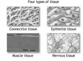
Based on structural and functional similarity, tissues are classified into four broad categories. Epithelial, connective, muscular, and neurological tissues are among them. The primary tissue types collaborate to contribute to the human body’s overall health and maintenance. As a result, any change in the structure of a tissue can result in injury or disease.
Classification of Tissues
Tissues are made up of cells with a similar structure and function. An organ is a collection of tissues that perform a specific function. In vertebrates, there are four distinct types of tissue: epithelial, connective, muscle, and nervous.
Epithelial Tissue
The skin and the linings of internal organs and body cavities are epithelial tissue, which consists of thin sheets of cells. Epithelial cells are densely packed, forming a protective barrier against injury, infection, and loss of water. Simple epithelium is a single layer of epithelial tissue; stratified epithelium is a multiple layer of epithelial tissue. In stratified epithelium, such as the skin, the damaged exterior cells are replaced by the division of cells beneath. Epithelial cells can be squamous (flattened), cuboid, or columnar in form. Certain epithelial tissues, such as the intestinal lining, absorb or emit chemicals.
Connective Tissue
Connective tissue is made up of cells embedded in an extracellular matrix and is classified as loose connective tissue, fibrous connective tissue, adipose (fat) tissue, cartilage, bone, and blood. Although connective tissue has a wide variety of characteristics, their primary function is to support and attach multiple tissues. Tendons, for example, are composed of fibrous connective tissue and serve to connect muscle to bone. The blood carries oxygen, nutrients, and waste products, as well as performing immune functions, in order to meet the needs of other tissues.
Muscle Tissue
Muscle tissue is made up of clusters of long, thin muscle cells referred to as muscle fibres. Muscle cells have the ability to contract and expand, which enables the body and its internal organs to move. The three major types of muscle tissue are cardiac muscle (found in the heart), skeletal muscle (found in bones, such as the limb muscles), and smooth muscle (found in visceral organs such as the intestines).
Nervous Tissue
Neurons—specialized cells that transmit, transport, and receive information via electrochemical signaling—and supporting cells called glial cells make up nervous tissue. A nerve is a collection of neurons. A brain is a collection of nerve cells. Apart from controlling muscle movement, nervous tissue detects sensory stimuli and directs a large number of the body’s activities.
Nervous tissue
The nervous system’s cells are designed to send electrical signals throughout the body. Nervous tissue contains two types of cells: neurons and glia.
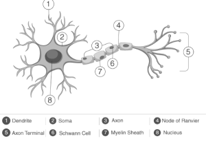
Neurons: Neurons have a big cell body called a soma and lengthy projections that transfer information. Axons or dendrites are the projections. Axons send outgoing impulses, while dendrites receive them. The axons of neurons are easily detected in longitudinal or cross-sectional slides. In the peripheral nervous system, ganglia are called nuclei in the central nervous system.
Glia: Glia outnumber neurons in nervous tissue. The nervous system’s cells vary. Astrocytes guard blood arteries and assist neurons near synapses. Oligodendrocytes are present in the brain’s white matter. Large projections from these cells wrap around a neuron’s axon insulating it for faster impulse projection.
Schwann cells do the same thing in the periphery. The sheathing provided by oligodendrocytes and Schwann cells helps identify nervous tissue. Microglia are the brain’s macrophages. These cells regularly scan the nerve tissue for intruders and trash.
Extracellular fluid transports ions and neuromediators to transfer impulses in nervous tissue. Glia regulate the extracellular environment because action potentials require a certain ion concentration. The blood brain barrier is formed by glia surrounding capillaries in nervous tissue.

Structure of Nervous Tissue
- It is constructed of nerve cells or neurons, each of which has an axon. Axons are long stem-like projections that emerge from the cell and are responsible for communicating with other cells known as Target cells and thereby transmitting impulses.
- The primary component of the cell is the nucleus, cytoplasm, and organelles. Processes are extensions of the cell membrane.
- A dendrite is a highly branching mechanism that receives information from neighbouring neurons and synapses (specialized point of contact). Dendrites transmit information from other neurons to the cell body via their connections.
- Information in a neuron is unidirectional, passing from dendrites to the cell body and down the axon.
Conclusion
Tissue is critical because it enables the study of disease progression, the determination of prognosis, and the identification of the most effective treatments for various diseases. It has made a significant contribution to the medical industry’s advancement.
 Profile
 Settings
 Refer your friends
 Sign out


