If you talk to a group of cell biologists about what they enjoy most about their jobs, you could discover that it all boils down to one thing: they’re all secretly microscope geeks. What they truly enjoy is sitting in a small, dark room for hours on end, communing with their preferred cell type through the lens of a magnificent microscope. That may seem strange, but cells, like living stained glass, may be rather beautiful.
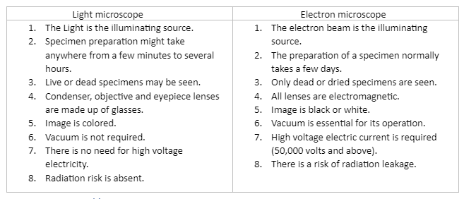
Light microscopes are the most common type of student microscope. A light microscope allows the user to see a magnified image by passing visible light through the specimen (the biological sample you’re looking at) and bending it via the lens system. Light microscopy has the advantage of being able to be done on living cells, allowing researchers to observe cells doing their usual functions (such as migrating or dividing) under the microscope.
Brightfield microscopes are common in student labs, which means that visible light passes through the sample and is immediately used to generate an image without any alterations. Light microscopy that is a little more advanced uses optical methods to boost contrast, making it easier to view features in cells and tissues.
Fluorescence microscopy is a type of light microscopy that is used to image samples that fluoresce (absorb one wavelength of light and emit another). The fluorescent molecules are excited by light of one wavelength, and the light of a different wavelength that they produce is collected and used to form a picture. Most of the time, the section of a cell or tissue we wish to examine isn’t inherently fluorescent and must be labelled with a fluorescent dye or tag before being examined under a microscope.
Beyond the approaches we outlined above, several cutting-edge types of light microscopy can provide extremely high-resolution images. However, if you want to view something very small at a very high resolution, you might want to try electron microscopy, which is a tried-and-true technique.
Electron microscopes differ from light microscopes in that they create an image of a specimen using a beam of electrons rather than light. Electron microscopes can produce higher-resolution images than ordinary light microscopes because electrons have a shorter wavelength than visible light. Electron microscopes can examine not just whole cells, but also cell components and compartments inside them.
However, in order for electron microscopy to work, materials must be placed under vacuum (and typically are prepared via an extensive fixation process). Because of this, live cells cannot be visualised.
Salmonella bacteria appear in a light micrograph (left) and an electron microscope image (right) (right). In the light microscope image, the bacteria appear as small purple dots, however in the electron micrograph, their shape and surface roughness, as well as characteristics of the human cells they’re attempting to infect, are plainly visible.
Electron microscopy is divided into two categories. A beam of electrons passes back and forth across the surface of a cell or tissue in scanning electron microscopy (SEM), providing a detailed image of the 3D surface. The image of the Salmonella bacteria at right, above, was taken using this technique of microscopy.
The material is cut into extremely thin slices (for example, using a diamond cutting edge) before imaging in transmission electron microscopy (TEM) and the electron beam passes through the slice rather than skimming over its surface. The transmission electron microscope (TEM) is frequently used to produce precise images of the internal architecture of cells.
Cells vary in size, although they’re usually pretty little. A typical human red blood cell, for example, is roughly eight micrometres in diameter (0.008 millimetres). To put things in perspective, a pin’s head is around one millimetre in diameter, so about 125 red blood cells might be lined up in a row across it. Individual cells, with a few exceptions, cannot be seen with the human eye, thus scientists must rely on microscopes.
A microscope is a device that magnifies items that would otherwise be too small to see, resulting in a picture that makes the object appear larger. The majority of cell photographs are taken with a microscope, and these images are referred to as micrographs.
The page contains all of the critical information that a student needs to know about the lens microscope and electron microscope at a basic level, such as its light microscope, electron microscope and difference between them, among other things. This is a vital piece of equipment for taking microscopes.
 Profile
 Settings
 Refer your friends
 Sign out