A Cockroach is probably the most loathed insect that ever existed. Cockroaches belong to the phylum Arthropoda. But the main fact is that cockroaches have inhabited the earth for the past 300 million years ago and still continue to be present on earth. Here, in this article we have discussed the external morphology and internal anatomy of a cockroach. The scientific name of the common cockroach is Periplaneta americana. The classification of cockroach is discussed below:
Phylum – Arthropoda
Class – Insecta
Order– Dictyoptera
Family– Blattidae
Genus –Periplaneta
Species– americana
Morphology of Cockroach
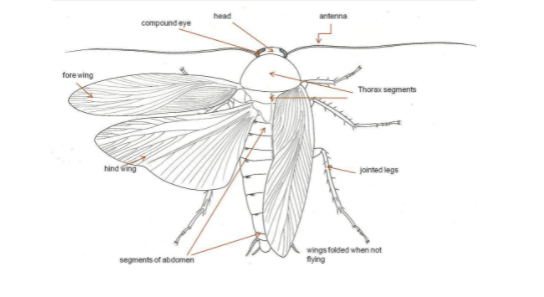
The body of the cockroach is generally segmented and elongated. They are dark brown or reddish brown in colour. Their exoskeleton is thick and hard composed of calcareous plates known as sclerites. There are about 10 segments. The segments present
— on dorsal side (or notum) are known as Tergum
—on the ventral side is known as Sternum.
The exoskeleton is generally coated with wax that is impermeable to water. This protects the body from loss of water and also helps in providing rigidity and surface for the attachment of body muscles. The adjacent segments are joined via a thin, soft and flexible membrane known as the arthrodial membrane.
The body is divisible into three regions such as head, thorax and abdomen.
A cockroach comprises three pairs of jointed appendages along with two pairs of wings.
The forewings of cockroaches are mainly mesothoracic and are commonly known as wing covers or tegmina or elytra. They are mainly used to cover the hindwings and have a protective function. Tegmina are dark, stiff , opaque and leathery in nature. The hind wings are usually large, thin, membranous and transparent. These wings are kept folded below the tegmina and possess the ability of flying.

Mouth Parts
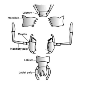
On the ventral side an opening known as mouth is present on the head which remains surrounded via different mouth parts mainly consisting of a pair of mandibles, first maxillae, labium or fused second maxillae, hypopharynx and labrum. These mouth parts of the cockroach help in ‘biting and chewing’ the food.
Functions of the mouth parts are discussed below:
Labrum: this is broad, flattened terminal sclerite present on the dorsal side of the head capsule, it is easily movable and articulated to the clypeus that acts as the upper lip. It possesses epipharynx ( that acts as a chemoreceptors) on its inner side.
Mandibles: these are thick hard and triangular appendages present beneath the labrum, on each lateral side of the mouth, that bears pointed, teeth like processes known as denticles.
First maxillae: these are located on each side of the mouth and next to mandibles suitable for cutting and chewing. They also contain olfactory receptors.
Labium: the labium or the second maxillae are fused together to form a single large structure that covers the mouth from ventral side, hence it is known as the ‘lower lip’ or labium. It also possesses tactile and gustatory sensory setae.
Hypopharynx: these are a small, cylindrical mouthpart, that is compressed between first maxillae and covered via labrum and labium on dorsal and ventral sides respectively. It also bears many sensory setae on its free end, and the opening of a common salivary duct upon its basal part.

Compound Eyes
At the dorsal surface of the head are located compound eyes. Each eye comprises about 2000 hexagonal ommatidia. These several ommatidia help a cockroach in receiving several images of an object. This kind of vision is commonly known as the mosaic vision. The image formed has more sensitivity and less resolution, and they possess nocturnal vision.
Legs of cockroach
The thorax of a cockroach attaches three pairs of legs. Each of the three pairs of legs was named after the region of the thorax to which it was attached:
The prothoracic legs are present very close to the cockroach’s head. These are the shortest legs, and they function like brakes during running. The middle legs are known as the mesothoracic legs. They help in moving back and forth. The longer metathoracic legs represent the cockroach’s back legs, and they help move the cockroach forward.
These three pairs of legs are generally different in lengths and functions, but they possess the same parts and move in the same way. The upper portion of the leg is known as the coxa and attaches the leg to the thorax. The other regions of leg includes:
The trochanter that acts like knees and helps the cockroach bend its leg.
The femur and tibia usually resemble thigh and shin bones.
The segmented tarsus resembles an ankle and foot. The hook-like tarsus provides cockroaches with the ability to climb the walls and walk upside down on ceilings.
Abdominal Segments
The abdomen is composed of 10 segments. The 7th, 8th and 9th sterna makes up the genital pouch in females. Whereas, in males, the genital pouch is located at the hind end of the abdomen. The male cockroach possess thread-like anal styles, that are absent in the female cockroaches. The 10th segment has a filamentous structure known as the anal cerci, in both male and female cockroach.
Anatomy of Cockroach
Alimentary Canal
The alimentary canal is divided into three; they are the foregut, midgut, and hindgut. The mouth ascends upto pharynx that further leads into a narrow passage known as the oesophagus. The oesophagus opens into a sac-like structure known as the crop that helps in storing food.
The gizzard represents the next structure which is present after the crop. It is also known as the proventriculus. Its main function is to grind the food particles due to the presence of six chitinous plates known as teeth. The entire foregut is lined by a cuticle. At the junction of the foregut and midgut, a ring of tubules known as the gastric caeca are present, that secretes digestive juice.
Malpighian tubules are another ring of 100-150 yellow coloured thin filamentous structures that are present at the junction of the midgut and hindgut. These tubules help in the removal of excretory products. The hindgut opens outside via the anus.
Circulatory System
Cockroaches possess an open blood vascular system since the blood vessels are poorly developed. There is an open space known as the hemocoel where the visceral organs are situated.
These visceral organs always bathed in hemolymph that represents the blood of a cockroach. The hemolymph is composed of a colourless plasma and haemocytes. An elongated tube that possesses muscular walls helps in regulating the blood in the hemocoel. This elongated tube which represents the heart of the cockroach possesses many funnel-shaped chambers and lies along the mid-dorsal line in the abdomen and thorax.
Respiratory System
The respiratory system of cockroaches possesses a network of trachea. They further open via 10 pairs of spiracles that are situated on the lateral side of the body. Thin tubes help in carrying oxygen from the air to all the other parts of the body. The spiracles can be regulated by the sphincters. Exchange of gases occurs via the process of diffusion.
Nervous System
Fused ganglia are segmentally arranged that make up the nervous system of cockroaches. The thorax region possesses three ganglia whereas the abdomen possesses six ganglia. In a cockroach, the nervous system is usually spread throughout the body.
The head region possesses only a little bit of the nervous system while the majority is located on the ventral side of the body. The supra-oesophageal ganglion helps in supplying the nerves to antennae and compound eyes. The sense organs in a cockroach are generally the antennae, eyes, maxillary palps, labial palps, anal cerci.
Excretory System
The Malpighian tubules help in the process of excretion in a cockroach. Glandular and ciliated cells are present lining each tubule, that absorbs the nitrogenous waste products. These are mainly converted into uric acid and are excreted out via the hindgut. It is because of this reason that a cockroach is known as uricotelic.
Reproductive System
The reproductive system of cockroaches is well developed in both the males and females. The male reproductive system possess a pair of testes that is located on the lateral side of the 4th -6th abdominal segments. An accessory reproductive gland is also present in the 6th and 7th abdominal segments and are generally mushroom shaped. Chitinous asymmetrical structures known as the male gonapophysis or phallomere comprises the external genitalia.
The female reproductive system possesses two large ovaries that are situated laterally in the 2nd to the 6th abdominal segments. Somewhat a group of eight ovarian tubules forms one ovary. They comprise a chain of developing ova. The fertilized eggs are encased in a casing known as the ootheca. Female cockroaches produce about 9 to 10 ootheca that contain around 14 to 16 eggs each.
Conclusion
About 300 million years ago cockroaches were found to occupy and will continue to be present on earth. So, here we come to an end of this topic. We hope that you were able to clear all the concepts regarding cockroaches.
 Profile
 Settings
 Refer your friends
 Sign out


