NEET UG » NEET UG Study Material » Biology » Difference between Epidermis and Dermis

Difference between Epidermis and Dermis

In this article, we study the difference between epidermis and dermis, Layers of Epidermis, Structure and Function, and more.

The body’s protective outer layers are the dermis and epidermis. The fundamental distinction between the dermis and the epidermis is that the dermis is a tissue beneath the epidermis that contains live cells, whereas the epidermis is the body’s outermost layer that protects it from dryness, damage, and infections. The dermis is a layer of dense irregular connective tissue present in the skin of animals. Animals have tiered layers of flattened cells in their epidermis. The dermis has blood vessels, but the epidermis has not. Plants have an epidermis as well. Plant epidermis has tightly packed cells with a waxy coating that prevents water loss.

Difference between Epidermis and Dermis

Parameter of Comparison

Epidermis

Dermis

Presence

The epidermis is the top layer of an organism’s skin, and it is made up of three layers.

The dermis is the skin layer that lies beneath the epidermis.

Blood Vessels

The epidermis is devoid of blood vessels.

The dermis contains blood vessels, which are a small network of blood capillaries.

Nerves

In the epidermis, nerves are not found.

In the dermis, there exist nerves.

Found

Epidermis is found on both plants and animals.

Dermis is a type of skin found only in animals.

Absorption of Nutrients

Diffusion from the dermis allows essential nutrients to be absorbed.

Through the blood capillaries, oxygen and critical nutrients are absorbed.

Layers of Epidermis

Stratum Basale: The stratum basale, also known as the stratum germinativum, is the deepest layer of the body, separated from the dermis by the foundation membrane (basal lamina) and linked to it by hemidesmosomes. This stratum contains cuboidal to columnar mitotically active stem cells that are constantly producing keratinocytes. 

Stratum spinosum: The 8-10 cell layers of the stratum spinosum, also known as the prickle cell layer, comprise irregular, polyhedral cells having cytoplasmic extensions known as “spines” that stretch outward and connect to neighbouring cells through desmosomes. Dendritic cells can be found in this stratum. 

Stratum granulosum: Diamond-shaped cells with keratohyalin granules and lamellar granules make up the 3-5 cell layers of the stratum granulosum. Keratin precursors can be detected in keratohyalin granules, which collect, crosslink, and form bundles over time. The lamellar granules contain the glycolipids that are released to the cell surface and act as a glue to connect the cells together.

Stratum lucidum: The stratum lucidum is a thin clear layer made up of eleidin, a keratohyalin transformation product present in thicker skin of the palms and soles.

Stratum corneum: The uppermost layer, the stratum corneum, is made up of keratin and horny scales made up of dead keratinocytes, also known as anucleate squamous cells. This layer’s thickness fluctuates the most, especially in calloused skin. Defensins, which are part of our first line of defense, are secreted by dead keratinocytes in this layer.

Structure and Functions

Structure and Function of Epidermis: Your skeletal system, organs, muscles, and tissues, as well as each layer of your skin, work together to keep your body safe. The epidermis also serves a variety of other purposes, including:

  • Hydration. The stratum corneum (outermost layer of the epidermis) stores water and maintains your skin hydrated and healthy
  • Skin cells are being created in large numbers. As you become older, new skin cells form at the bottom layer of your epidermis (stratum basale) and move up through the other layers. They reach the epidermis’ outermost layer, where skin cells shed from your body and new cells sprout at the bottom layer, after about a month
  • Protection. The epidermis protects your body from UV radiation, pathogens (bacteria, viruses, fungi, and parasites), and chemicals by acting as a shield
  • Color of the skin. Melanocytes are cells in the epidermis that produce melanin, a group of pigments that gives your skin its colour.


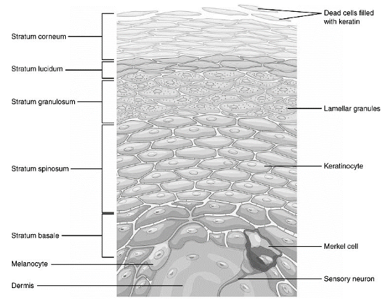
Structure and Function of Dermis

  • Reticular dermis: The reticular layer is the basal layer of the dermis. It is thick and contains blood arteries, glands, hair follicles, lymphatics, nerves, and fat cells. A net-like arrangement of elastin and collagen fibers surrounds the reticular dermis. These fibers help to sustain your skin’s overall structure while also allowing it to move and stretch
  • Papillary dermis: The top layer of your dermis is called the papillary layer. It is significantly thinner than the reticular dermis. Collagen fibers, fibroblast cells, fat cells, blood arteries (capillary loops), nerve fibers, touch receptors (Meissner corpuscles), and bacteria-fighting cells make up this structure (phagocytes). The papillary dermis extends to the epidermis’s basement layer. They develop a tight bond that unites them like fingers interlocking

Conclusion

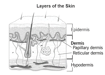
The top two layers of skin on your body are the dermis and epidermis. The top layer is your epidermis, while the intermediate layer is your dermis. Between your epidermis and hypodermis is your dermis.The epidermis is the skin’s thinnest layer. It aids in the hydration of your body, the production of new skin cells, the protection of your body from harm, and the production of melanin, which gives your skin colour.The epidermis is the thinnest layer of skin, whereas the dermis is the thickest. Collagen and elastin, which help make your dermis thick and supportive of your skin’s general structure, are found in your dermis.Your dermis contains all of your connective tissues, nerve endings, sweat glands, oil glands, and hair follicles.

faq

Frequently asked questions

Get answers to the most common queries related to the NEET UG Examination Preparation.

What is the dermis made up of?

Ans: Collagen ...Read full

What are the most prevalent indications and symptoms of dermis disorders?

Ans: Arteries, arterioles, capillaries, ...Read full

What are some of the most popular tests used to determine the health of the dermis?

Ans: Genetic testing: Looking for a def...Read full

What are the most frequent dermis treatments?

Ans: Corticosteroids. Inflammation and tissue damage are reduced with corticos...Read full