A microscope is an optical device that magnifies objects that are too small to be seen with the naked eye. Microorganisms are invisible to the naked eye, but they can be seen under a microscope. The microscope magnifies these micro-objects, allowing the user to view small details at a scale that is suitable for research and analysis.
Microscopy is the study of objects that are too small to be seen with the naked eye using microscopes. This article will explain the history of microscopes, as well as the various types and applications of microscopes.
Simple and compound microscopes differ in that compound microscopes have a much higher magnification capability and are employed in more intensive study. Stereo, inverted, metallurgical, and polarising microscopes are some of the different microscopes available.
Microscopy
It is made up of two Greek words: micros, which means small, and scope, which means to look at or see. Microscopy is a field of science that uses microscopes to examine cells and tissues that are not visible to the human eye.
Compound Microscope
Dual-lens microscopes are known as compound microscopes. The objective lens and the eyepiece are the two lenses. A student microscope is how most people refer to it.
A compound microscope serves the same goal as a simple microscope. It magnifies real-world objects that are microscopic to the naked eye using numerous lenses. It is used for professional applications that necessitate extensive investigation. On one side, it has a flat mirror surface, while on the other, it has a concave mirror surface. Multiple lenses are used in compound microscopes.
Lens used: Two convex lenses are used in a compound microscope, one in the eyepiece and the other in the objective. The magnifications of the eyepiece lenses vary depending on their focal lengths. A student microscope has three objective lenses with magnification powers of 10x, 40x, and 100x, and an eyepiece with magnification powers of 5x, 10x, 15x, 20x, and 30x.
Principle: The objective lens collects light scattered from the specimen, whereas the eyepiece is utilised to view the image in its entirety. It creates a simulated, erect, and enlarged image.
Magnification: The magnifying power of a lens is proportional to its focal length, and it is usually more than that of a simple microscope.

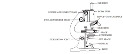
Parts of a Compound Microscopes
There are Following Parts of a compound microscope which are mention below:
Head/Body
It’s the component of the microscope that’s on top. The following components are housed in the microscope’s body:
Eyepiece
An ocular lens is another name for it. It is the lens that is located at the metal tube’s top end. Binocular models have two tubes, each with one eyepiece that can be changed in distance. Monocular models have a single tube, but binocular versions have two tubes, each with one eyepiece that can be adjusted in distance.
Eyepiece tube
This is a 160mm (6.3 inch) long metal tube. This measurement is based on the human eye’s resolution and is intended to reduce aberrations.
Objective lens
These are the microscope’s primary lenses.
Nosepiece
It retains the objective lenses and rotates to allow for the selection of a certain objective to face the specimen.
Coarse and Fine Focus Knobs
These are used to focus the image by moving the body tube. Coarse focus knobs are used to do rough image focusing, whereas fine focus knobs are used to remove any fuzzy focus and are entirely dependent on the eye’s resolution power.
Stage
A flat platform onto which a specimen is mounted. Because this is a mechanical stage, it is possible to shift it slightly for greater illumination.
Stage clips
Use finger movements to see different sections of the specimen while holding the slide firmly in place.
Aperture
The opening via which light is transferred to the stage and, in turn, to the specimen.
Iris Diaphragm
This is necessary because different specimens require varying amounts of light in order to achieve crisp contrasts and a higher resolution image. As a result, the iris diaphragm aids in improving contrast by regulating the amount of light.
Base
The base of the microscope is where the illuminator or mirror is kept.
Illuminator: A low-voltage halogen bulb is frequently present. Many models, however, employ natural light that is reflected back into the aperture.
Arm
The arm is the connecting piece between the upper and lower components of the microscope, and it is used to pick up and move microscopes. The arm and base are joined by a limited-movement inclination joint that allows the lighting and light through the specimen to be adjusted.
Working Procedure of Compound Microscope
There are following steps:
- A biological specimen is mounted on a clear glass slide that has been dipped in glycerine or water and covered with a coverslip.
- The prepared slide is then placed between the condenser and the objective lens on the stage.
- A reflector mirror and condenser focus a beam of visible light on the specimen.
- The amount of light can also be controlled by adjusting the iris aperture.
- Depending on the magnification necessary, an objective lens (fixed to the revolving nosepiece) is selected. The specimen must be immersed in oil when using a 100x objective lens.
- The objective lens collects light passing through the material, which an observer can see through the eyepiece.
- A coarse and fine adjustment knob can be utilised to get a clear image based on the magnification and resolution of the image.
- If oil immersion is utilised, a cotton cloth should be used to wipe the objective lens, specimen, and stage clean.
Application of Compound Microscope
- It’s most commonly used in pathology labs to identify viruses and bacteria.
- This microscope can be used to examine plant cells and the bacteria that live on them.
- It is used to determine the presence or absence of minerals and metals in blood samples and other biological materials.
- It aids in the examination and comprehension of the microbiological field of viruses and bacteria, which is impossible to observe with the naked eye.
- It’s utilised in a forensics lab to aid in the investigation of crimes.
- It’s even utilised to perform academic experiments at schools and colleges.
Significance of Microscopy
There are following Importance of Microscopy:
- In biology and medicine, microscopy offers a wide range of uses.
- It is mostly utilised to understand tissue and cellular architecture.
- Various types of microscopes are used to investigate subcellular structures.
- Infrared microscopes are used to determine whether two samples are identical.
- UV microscopy is particularly useful for studying protein crystal formation.
- Live specimens can be observed using dark field microscopy.
- When 3D structures are important, confocal microscopy is used.
- Single-molecule signals are detected using fluorescence microscopy.
Conclusion
A microscope is an optical device that magnifies objects that are too small to be seen with the naked eye. Microorganisms are invisible to the naked eye, but they can be seen under a microscope.A compound microscope serves the same goal as a simple microscope. It magnifies real-world objects that are microscopic to the naked eye using numerous lenses. It is used for professional applications that necessitate extensive investigation. On one side, it has a flat mirror surface, while on the other, it has a concave mirror surface. Multiple lenses are used in compound microscopes.
Application of Compound Microscope
- It’s most commonly used in pathology labs to identify viruses and bacteria.
- This microscope can be used to examine plant cells and the bacteria that live on them.
- It is used to determine the presence or absence of minerals and metals in blood samples and other biological materials.
 Profile
 Settings
 Refer your friends
 Sign out


