Coat protein complexes, Rab GTPases, tethering factors, and membrane fusion catalysts are required for vesicular transport of protein and lipid cargo from the endoplasmic reticulum (ER) to cis-Golgi compartments. Cargo is delivered by ER-derived vesicles to an ER-Golgi intermediate compartment (ERGIC), which then fuses with and/or matures into cis-Golgi compartments.
A retrograde directed pathway that recycles transport machinery back to the ER balances the forward transport pathway to cis-Golgi compartments. It is unclear how trafficking through the ERGIC and cis-Golgi is coordinated to maintain organelle structure and function, which raises important questions about trafficking routes and the organisation of the early secretory pathway.
What is Cis Golgi Network?
The cis-Golgi network is a large tubulovesicular network that connects to the cis face of the Golgi stack and receives and processes biosynthetic output from the ER.
Structure
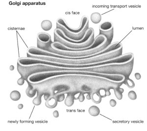
The golgi is composed of 5-8 folds known as cisternae. The cisternae contain specific enzymes that form five functional regions that modify proteins that pass through them in a stereotypical manner, as shown below:
- Cis-Golgi network: The cis-Golgi network faces the nucleus, connects to the endoplasmic reticulum, and serves as the entry point into the Golgi apparatus.
- Cis-Golgi: a large processing area that allows for biochemical modifications.
- Medial-Golgi: The medial-Golgi is a large processing area that allows for biochemical modifications.
- Trans-Golgi: a large processing area that allows for biochemical modifications.
- Trans-Golgi network: exit point for vesicles budding off the Golgi surface, where biochemicals are packaged and sorted according to their destination.

What is the Cis-face of Golgi apparatus?
Cis face is one of three Golgi apparatus networks, the others being the cis Golgi network (CGN), the medial compartment, and the trans-Golgi network (TGN). The cis face of the Golgi apparatus’s primary function is to receive proteins and lipids from the endoplasmic reticulum. As a result, the CGN is always in contact with the endoplasmic reticulum.
Because the cis face receives vesicles, it is always referred to as the forming face. It is also the first stage of substance packaging.
What is the trans-face of the Golgi apparatus?
The trans face, also known as the TGN, is the Golgi apparatus’s final phase. TGN’s primary function is to produce vesicles containing mature proteins or lipids. Immature proteins and lipids migrate to the Golgi apparatus’s medial compartment to mature. These substances are modified in a variety of ways, including post-translational modifications, glycosylation, and phosphorylation.
Exocytotic vesicles, secretory vesicles, and lysosomal vesicles are the three types of vesicles that leave the trans face of the Golgi apparatus.
Exocytotic vesicles contain proteins that are released extracellularly, such as antibodies, whereas secretory vesicles contain substances that are released extracellularly, such as neurotransmitters. Furthermore, lysosomes contain digestive enzymes involved in phagocytosis as well as membrane proteins that will fuse with the membrane.
Golgi Apparatus
The Golgi apparatus (GA), also known as the Golgi body or the Golgi complex and found in both plant and animal cells, is made up of a series of five to eight cup-shaped, membrane-covered sacs called cisternae that resemble a stack of deflated balloons. However, in some unicellular flagellates, up to 60 cisternae may join together to form the Golgi apparatus. Likewise, the number of Golgi bodies in a cell varies with its function. Animal cells typically have ten to twenty Golgi stacks per cell, which are linked together by tubular connections between cisternae to form a single complex.
This complex is usually found near the cell nucleus.The Golgi apparatus was one of the first organelles to be discovered due to its relatively large size. In 1897, an Italian physician named Camillo Golgi was studying the nervous system with a new staining technique he developed (and which is still sometimes used today; known as Golgi staining or Golgi impregnation), when he noticed a cellular structure he called the internal reticular apparatus in a sample under his light microscope.
The structure was named after him soon after he publicly announced his discovery in 1898, and it became known as the Golgi apparatus. Many scientists, however, did not believe that what Golgi saw was a real organelle in the cell, instead claiming that the apparent body was a visual distortion caused by staining. The Golgi apparatus is a cellular organelle, according to the electron microscope, which was invented in the twentieth century. Proteins, carbohydrates, phospholipids, and other molecules formed in the endoplasmic reticulum are transported to the Golgi apparatus and biochemically modified as they move from the complex’s cis to trans poles.
Conclusion
The cis-Golgi network is a large tubulovesicular network that connects to the cis face of the Golgi stack and receives and processes biosynthetic output from the ER. It is unclear how trafficking through the ERGIC and cis-Golgi is coordinated to maintain organelle structure and function, which raises important questions about trafficking routes and the organisation of the early secretory pathway.The cisternae contain specific enzymes that form five functional regions that modify proteins that pass through them in a stereotypical manner.: The cis-Golgi network faces the nucleus, connects to the endoplasmic reticulum, and serves as the entry point into the Golgi apparatus. The Golgi apparatus , also known as the Golgi body or the Golgi complex and found in both plant and animal cells, is made up of a series of five to eight cup-shaped, membrane-covered sacs called cisternae that resemble a stack of deflated balloons.
 Profile
 Settings
 Refer your friends
 Sign out


