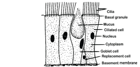
Several columnar or cuboidal cells with hairlike appendages that may beat quickly form a piece of epithelium (see cilium). The ciliated epithelium is responsible for conveying particles or fluid through the epithelial surface of structures such as the trachea, bronchial tubes, and nasal cavities. It is usually seen in the vicinity of goblet cells, which secrete mucus.
Cilia are little hair-like protuberances that protrude from the surface of eukaryotic cells (or cilia in plural). They are in control of the motility of the cell itself as well as the fluids that are present on the cell surface. They also play a role in the perception of mechanical forces. They have also given rise to a type of bacteria that are based on these small structures. A ciliate is a protozoan that has cilia on its body, which it uses for both locomotion and digestion.
Ciliated epithelium
A columnar or cuboidal epithelial region with hairlike appendages (cilium) capable of rapid beating. For example, the trachea, bronchial tubes, and bronchioles all include ciliated epithelium, which serves the function of transporting particles or fluid over their epithelial surfaces. Nostrils and nasal cavities are frequently seen in the proximity of goblet cells, which secrete mucus.
Location
The pulmonary system, including the trachea and bronchi, is covered by ciliated columnar epithelial cells, which are spread throughout the body. They’re also found in the fallopian tubes, which are part of the female reproductive system.
Function
The goblet cells are usually mixed with a simple ciliated epithelial cell, which may be found in the pulmonary system, and this is their function. Mucus is produced to create a mucosal layer apical to the epithelial layer, which is surrounded by the epithelial layer. The rowing-like action of epithelial cilia, in conjunction with the movement of goblet cells, is always effective in driving mucus from the lungs.
Infection caused by particulate particles can be prevented with the usage of this product. In a 2002 research, Lawson came to the conclusion that ciliated cells are crucial in the healing of distal airway damage. This population of pulmonary epithelial cells is believed to have reached the end of their differentiation and will never divide again. When the bronchiolar epithelium is healed after tissue injury, park (2006) discovered that the ciliated cells undergo morphological alterations, changing their shape from squamous to cuboidal to columnar forms, showing that they have the capacity for differentiation.
Diagram

Uses of Epithelium Cells
Some structural and functional traits are shared by all epithelia, while others are unique to each. There is little or no extracellular material between the cells in this tissue, which results in a tightly packed cell structure. Desmosomal junctions and tight junctions are types of cell junctions that connect two or more neighbouring cells. In this case, the apical surface of epithelial tissue is visible, whilst the basal surface serves as an anchoring layer, connecting the epithelial tissue to the underlying connective tissue. In order to adhere to connective tissue, the basement membrane, which is composed of proteins, must first bind to connective tissue.
Cilia are tiny hair-like protuberances that protrude from the exterior of eukaryotic cells (cilia are used when the word cilium is plural). They are in charge of the movement of the cell itself as well as the movement of the fluids on the cell surface. They are also involved in the perception of mechanical forces. Also named after these little structures is a group of bacteria that exist in the environment. Ciliates are protozoans that have cilia on their bodies, which they employ for both movement and eating.
Conclusion
Ciliated columnar epithelium is made up of simple columnar epithelial cells that have cilia on the apical surfaces of their cellular surfaces. They may be found in the lining of fallopian tubes as well as other sections of the respiratory system, where the beating of cilia aids in the removal of dust and other particulate matter. The majority of these tissues are made up of goblet-shaped cells that release mucus and longer columnar cells that are not mucus producing. It travels the full length of the epithelium. They also comprise short basal cells, the apical surfaces of which never reach the lumen of the cell body. Basal cells have the potential to develop into goblet cells or columnar cells.
 Profile
 Settings
 Refer your friends
 Sign out


