Profile
 Settings
 Refer your friends
 Sign out


The health of a plant is inextricably linked to its roots. When roots become weak or infected, the entire plant suffers. In order to be healthy, the roots must continue to expand.
The root system performs several critical activities. The roots are responsible for absorbing the water and nutrients that a plant requires to survive. The roots attach the plant to the ground and provide support to the plant’s above-ground parts. The photosynthesis-produced food is stored in the roots. This food can be utilised at a later time when a plant requires it for growth or survival. As root performs such important functions, it is very important to know the anatomy of dicot root and monocot root to understand functions of internal structures of monocot and dicot roots. The anatomy of monocot and dicot roots is an important topic in biology that students must prepare thoroughly.
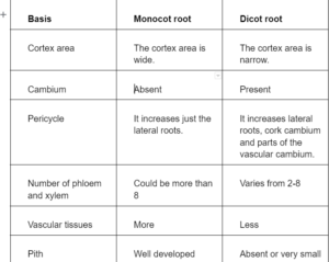
The internal structure of the dicot root and monocot root-
Examples of dicot plant-Gram, Pea, Pulses,Beans,Peanut etc.
Examples of monocot plants-Maize,Rice,Banana,Sugarcane etc.
The internal structure of the root consists of epiblema, cortex, endodermis, pericycle, conjunctive bundles and vascular tissues.
Epiblema
- It is also known as the piliferous layer or rhizodermis
- It is the root’s outermost single layer, made up of thin-walled, tightly packed parenchymatous cells with no intercellular gaps
- The stomata and cuticle are missing
- Most epidermal cells protrude from the epidermis in tubular unicellular root hairs
- The piliferous layer is called for the root hairs in epiblema
- This layer has no cuticle and is responsible for absorbing mineral salts and water from the soil
- The surface area available for absorption is maximised by root hairs
Cortex
- It is simpler and more uniform, generating a huge zone made up of unspecialised parenchyma cells with visible intercellular gaps. The cells are alive and have a lot of leucoplasts
- It is primarily concerned with food storage. However, at an early stage, it is in charge of water and solute translocation to conducting components. The endodermis is the last layer of the cortex
- It is found only in roots and is made up of tightly packed barrel-shaped cells that create a unique zone around the stele. The radial walls of endodermal cells have Casparian thickenings
Endodermis
- It is the deepest layer, consisting of a single layer of closely packed parenchymatous cells with no intercellular gaps
- This layer’s radial walls are frequently thickened, and this thickening can occasionally extend to the inner walls as well
- The thickening is caused by the deposition of suberin and lignin
- Strip or band-like formations called Casparian strips or Casparian bands occur as a result of deposition
- Thin-walled endodermis cells placed opposite proto-xylem components are referred to as passage cells because they allow water to travel from the roots to the xylem
- Around the stele, the endodermis serves as a waterproof jacket
Pericycle
- The pericycle is a layer of cells located immediately inside the endodermis. Because of its meristematic capabilities, the pericycle is significant. It is from here that lateral (branch) roots emerge
- Before penetrating the soil, the lateral roots push their way through the other layers of cells. Woody plant roots’ secondary (woody) development is also influenced by the pericycle
Conjunctive bundles
- Between xylem and phloem bundles, there are one or many layers of thin elongated parenchymatous cells that have no intercellular spaces. These constitute conjunctive tissue
- It is responsible for storing food
Vascular tissues
- The vascular tissue of dicot roots is found in the middle of the root. Inside the pericycle, vascular tissue may be seen near the root’s core. Xylem tissue is located in the heart of a dicot root and looks like a star or an X in cross-section. An arm is generally where lateral roots begin to form. Between the arms of the star is phloem tissue, which transports carbohydrates to the roots. Between the xylem and the phloem is the vascular cambium
- Monocots have root tissues that are comparable to dicots. The epidermis, cortex, endosperm, and pericycle are all similar in appearance. The vascular tissues, on the other hand, are organised differently. Monocots’ circulatory tissues form bundles, with the xylem on the inside. The bundles create a ring around the centrally placed pitch
The internal structure of the dicot root
Epiblema is the outermost layer, and epiblema cells appear in the form of unicellular root hairs. Multiple layers of thin-walled parenchyma cells make up the cortex. The endodermis is the cortex’s deepest layer, made up of barrel-shaped cells with no intercellular space. Endodermal cells have a waxy covering on their tangential and radial walls known as Casparian strips.
The pericycle, which comprises layers of thick-walled parenchymatous cells, is located next to the endodermis. During secondary growth, the beginning of vascular cambium and lateral roots takes place in these cells. Conjunctive tissues are the parenchymatous cells that lie between the phloem and the xylem. It has two to four patches of xylem and phloem. The stele is made up of all tissues found inside the endodermis, such as the pericycle, vascular bundles, and pith.
The internal structure of the monocot root
In many ways, the anatomy of a monocot root is nearly identical to that of a dicot root. Cortex, epidermis, endodermis, pericycle, pith, and vascular bundles are also present. The total number of xylem bundles is the difference. Monocot roots often have six xylem (polyarch) bundles more in comparison to dicot roots, which have fewer. Monocotyledonous roots don’t experience secondary growth and have a well-developed pitch.
Conclusion
The internal structure of roots plays an important role in the sustenance of plant life. Internal structures of monocot roots and dicot roots are almost similar, with a few differences here and there.