 Profile
 Settings
 Refer your friends
 Sign out


The inner lining of mucosa, the submucosa containing blood vessels and lymphatics, the muscularis externa or smooth muscle layer, and the outermost layer or serosa/adventitia make up the GI tract’s organs. Each tissue layer has a specific function in the digestive system, ranging from forming a protective barrier against the stomach’s highly acidic contents to delivering hormones, creating muscle contractions, and draining lymph. Supporting cells, for example, are present in the epithelium lining the inside of the stomach and create a protective layer of mucus and gastric acid for digestion. Other supporting cells, such as the stomach’s mucosal cells and the pancreas’ acinar cells, also generate zymogens, inactive forms of digestive enzymes that are stimulated to become active enzymes.
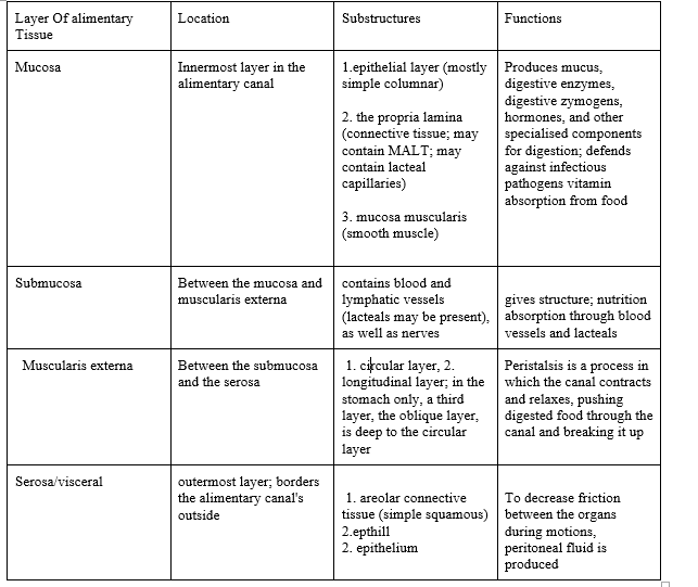
MALT = mucosa-associated lymphatic tissue; immune functions
lacteal = fat-absorbing lymphatic vessels in the small intestine
1The primary diagnostic features of the tubular digestive tract’s components are highlighted in these drawings. Except for the small intestine, which is depicted in the two smaller panels in the lower left quadrant, all photos show tubular digestive organs cut in the cross section. The longitudinal sections of the small intestine are depicted in these sections. Pink mucosae, orange submucosa, red muscularis externa, and blue-grey serosa / adventitia.
2.Stratified squamous moist epithelium lines the oesophagus. The upper and lower oesophagus’ lamina propria, as well as the submucosa, have scattered glands. Muscularis externa starts off as skeletal muscle, then becomes skeletal plus smooth muscle, and lastly smooth muscle alone in the bottom half. The organ is surrounded by an adventitia.
3.The stomach has a thick mucosa that is bordered with a surface sheet gland. Stomach glands line the lamina propria, each one opening into a gastric pit. The muscularis externa is extremely thick, with three subdivisions rather than the usual two. A serosa protects the stomach.
4.Small intestine villi are conspicuous features. The glands of the intestine open at the bases of the villi and extend to the muscularis mucosae. The duodenum is distinguished from the rest of the small intestine by glands in the submucosa. Although a section of the duodenum is retroperitoneal and has an adventitia, the organ is mostly covered by a serosa.
5.The large intestine (colon) lacks villi and has lamina propria with very straight intestinal glands. There are no submucosal glands present. The taenia coli are three longitudinal strips that make up the majority of the muscularis externa outer longitudinal subdivision. Depending on its location, the large intestine is covered with an adventitia or a serosa.
Conclusion
We conclude that the alimentary canal is the passageway via which food enters our bodies and exits via the anus after digestion. It is a tube-like structure that extends from the mouth to the anus. The alimentary canal, often known as the digestive tract, is a vital part of human digestion.


