Mitochondria serve as energy-producing powerhouses, they are abundant in places where energy is required in large quantities, such as the sperm tail, muscle cell, liver cell (which can contain up to 1600 mitochondria), microvilli, and the oocyte (which can contain more than 300,000 mitochondria).
In most cells, there are approximately 2000 mitochondria per cell, which accounts for approximately 25% of the total cell volume.
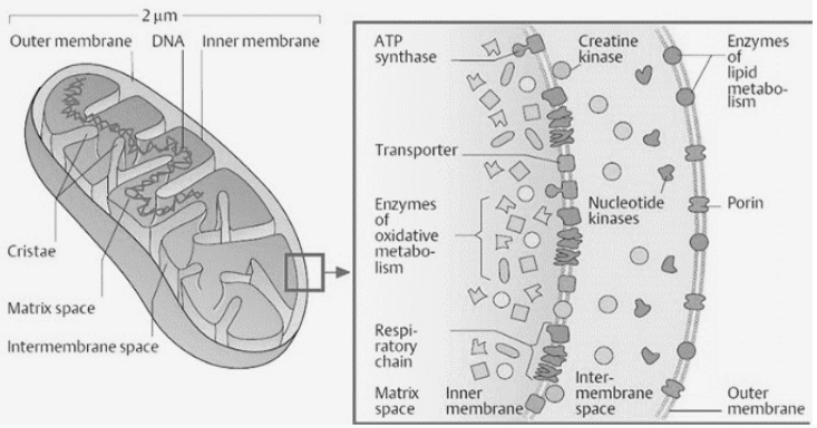
Richard Altmann published the first description of mitochondria in 1890, referring to them as “bioblasts.” Benda first used the term “mitochondria” in 1897, when he published a paper on the subject. Mitochondria are organelles that are mobile and plastic, and they have a double-membrane structure. It has a diameter that ranges from 0.5 to 1.0 micrometer. It is divided into four different contexts: the outer membrane, the inner membrane, the intermembrane space, and the matrix.
It is a viscous or gel-like fluid that consists of a mixture of enzymes, ribosomes, inorganic ions, mitochondrial, nucleotide coagulation factors, and organic molecules. It is involved in the process of cellular respiration as well as the generation of ATP molecules in the body.
Cristae is a term used to refer to the inner layer of the mitochondrial matrix that is surrounded by folds in the mitochondrial matrix. In addition to increasing the surface area of the inner membrane.
The mitochondrial ribosome, also known as the mitoribosome, is a type of ribosome that is found within the mitochondria. It is a protein complex that performs its function by interpreting mitochondrial mRNAs encrypted in mitochondrial DNA (mtDNA).
The inner mitochondrial membrane contains proteins and performs its function by allowing only specific molecules to enter and exit the cell. As a result, they are also referred to as “special membrane transporters” (SMT).
Porins are proteins found in the outermost layer of mitochondria that act as channels, allowing proteins to move freely between the inner and outer membranes of the mitochondrial cell. It also contains a large number of enzymes that perform a wide range of functions.
The space between the inner and outer membranes is referred to as the interstitial space. Each of the two sub compartments, the intercristal space and the lumen, is subdivided into a separate compartment. The cristae junctions, which are tubular structures ranging from 10 to 40nm in diameter, are what separate the two.
Endosymbiotic and autogenous origins of mitochondria are the two hypotheses that have been advanced in recent years. In accordance with this hypothesis, mitochondria were initially prokaryotic cells, involved in executing oxidative processes that were not entirely feasible for eukaryotic cells; they then evolved into endosymbionts that lived inside the eukaryote and shared its metabolic processes. Autogenous mitochondria, according to the autogenous hypothesis, were formed by a component of DNA from the nucleoplasm of the eukaryotic cell at the period of deviation with the prokaryotes; this DNA component would’ve been embedded by membranes, which prevented proteins from crossing it. As a result of the fact that mitochondria share many characteristics with bacteria, the endosymbiotic hypothesis is more broadly acknowledged.



 Profile
 Settings
 Refer your friends
 Sign out